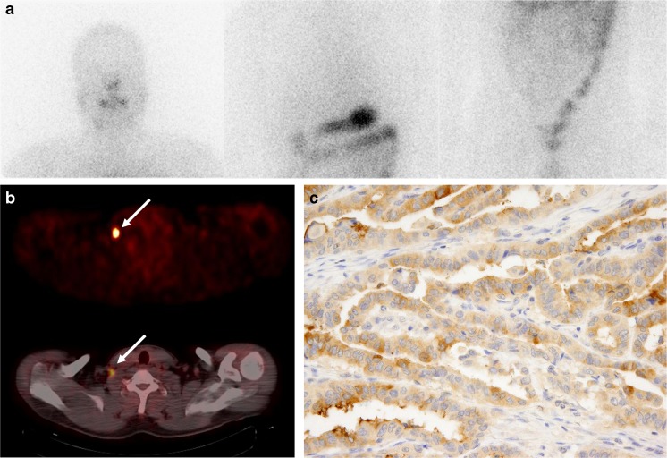
Fig. 1.
Anterior post-therapeutic 131I scan image (a) and 18F-FDG PET and fused PET/CT images (b) of a 42-year-old male patient with papillary thyroid cancer. 18F-FDG PET and PET/CT images (b) showing focal intense 18F-FDG uptake in the right lower neck lymph node (arrow), whereas the 131I scan image (a) shows no abnormal 131I uptake. Immunostaining for thyroglobulin (Tg) in the surgical specimen of the right lower neck node (c) showed weak Tg expression

