Fig. 1.
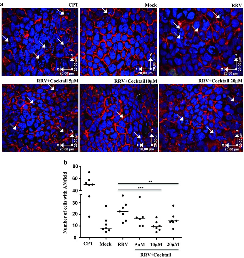
Treatment of Caco-2 cells with caspase inhibitors during RV infection decreases apoptotic nuclei. Caco-2 cells cultured on microscope slides pretreated with collagen in DMEM supplemented with 20 % FBS for 10 days were treated with caspase inhibitors (z-VAD-fmk, z-DEVD-fmk, and z-LEHD-fmk) in a cocktail at different concentrations for 1 h and then infected for 45 min with the RRV strain (moi of five). The inhibitors were maintained during the virus inoculation and 18 h after infection. As a control of apoptosis induction, we use camptothecin (CPT). a Visualization of apoptotic nuclei (white arrows) in a representative image of seven independent experiments (nuclei dyed with DAPI (blue) and filamentous actin with phalloidin Alexa-fluor 594 (red)). The images were acquired with an Olympus FV-1000 confocal microscope. b Number of cells with apoptotic nuclei by field. The median of seven independent experiments is shown. Each single point represents the median of counting six different optical fields. The statistical significances are shown using non-parametric test for paired or non-paired data (Wilcoxon or Mann Whitney tests p < 0.05*, p < 0.01**, p < 0.001***)

