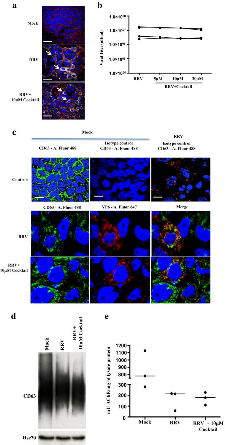
Fig. 2.
The presence of caspase inhibitors during RV infection does not affect viral replication or the intracellular expression of CD63 and Hsc70 or AChE activity. Caco-2 cells cultivated as described were treated with the caspase inhibitor cocktail (10 μM de z-VAD-fmk, z-DEVD-fmk, and z-LEHD-fmk) for 1 h and then infected for 45 min with RRV (moi of five) or the control mock infection. The inhibitors were maintained during the virus inoculation and 18 h after the infection. a Cell nuclei were stained with DAPI (blue), filamentous actin were detected using phalloidin Alexa-fluor 594 (red), and VP6 was identified by using a Mab anti-VP6 followed by a mouse anti-IgG polyclonal antibody conjugated with Alexa-fluor 647 (gray). White thick lines correspond to 40 μm. Arrows show infected cells b The viral particles present in the supernatants of the treated cells were subjected to a viral titration assay by immunocytochemistry on MA104 cells. Each point represents the viral titter obtained for each condition. Results from 4 independent experiments are shown; points corresponding to the same experiment are connected by lines. c Cell nuclei were stained with DAPI (blue), and viral protein VP6 was identified using a Mab anti-VP6 followed by a mouse anti-IgG polyclonal antibody conjugated with Alexa-fluor 647 (red), and Mab anti-CD63 conjugated with FITC (in green). As an isotype control, an IgG1κ Mab was conjugated with FITC. A representative image of three independent experiments is shown. White thick lines correspond to 20 μm. d WB evaluation of CD63 and Hsc70 from cell lysates (20 μg of protein per row). A representative image of three independent experiments e AChE quantification showing the quantity of mU of AChE activity present in each mg of protein from samples of three independent experiments

