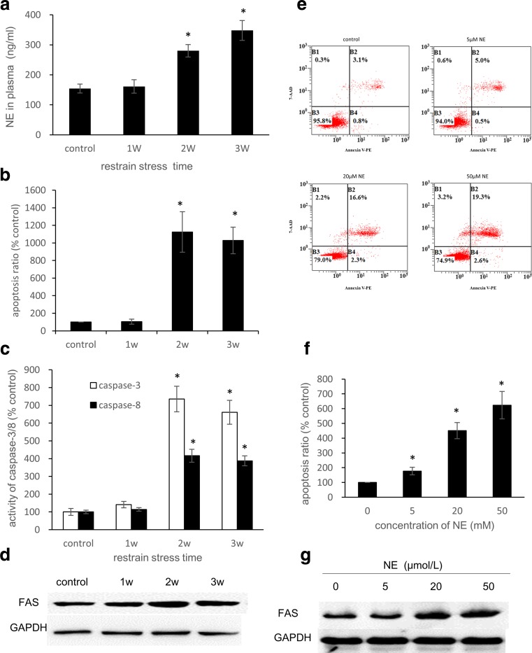
Fig. 1.
Restraint stress induces apoptosis in rat myocardia, and NE treatment induces apoptosis in H9C2 cells. a Plasma NE levels of the rats after restraint stress. The rat plasma NE levels were measured by ELISA (n = 3). b In the TUNEL assay, the apoptosis ratio was defined as the ratio of positive cells to the total number of cells in each visual field. In each section, five visual fields were counted and averaged (n = 5). c Caspase-3 and Caspase-8 Activity Assay Kits were used to detect caspase-3/8 activity. e, f After 36 h of treatment with NE at concentrations of 0, 5, 20, and 50 μM, H9C2 cells were stained with 7-AAD and Annexin V-PE and analyzed by FCM. d, g Whole-cell lysates were subjected to western blot analysis using the anti-FAS antibody. GAPDH was used as a loading control. The bars represent the mean ± SD of three experiments performed in triplicate. *P < 0.05 vs the control group

