Fig. 3.
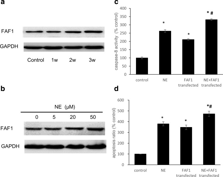
FAF1 levels increased with stress intensity, and overexpression of FAF1 exacerbated stress-induced apoptosis. a After restraint stress, whole-cell lysates of rat myocardia were subjected to western blot analysis using an anti-FAF1 antibody. GAPDH was used as a loading control. b Levels of FAF1 in H9C2 cells treated with different concentrations of NE for 36 h. c After transfection with the pcDNA3.1(+)-rFAF1 plasmid, the activity of caspase-8 in whole-cell lysates was detected using a Caspase-8 Activity Assay Kit. d Flow cytometric analysis of apoptosis. The data shown represent the mean ± SD from three independent experiments. *P < 0.05 vs the control group. #P < 0.05 vs the NE group

