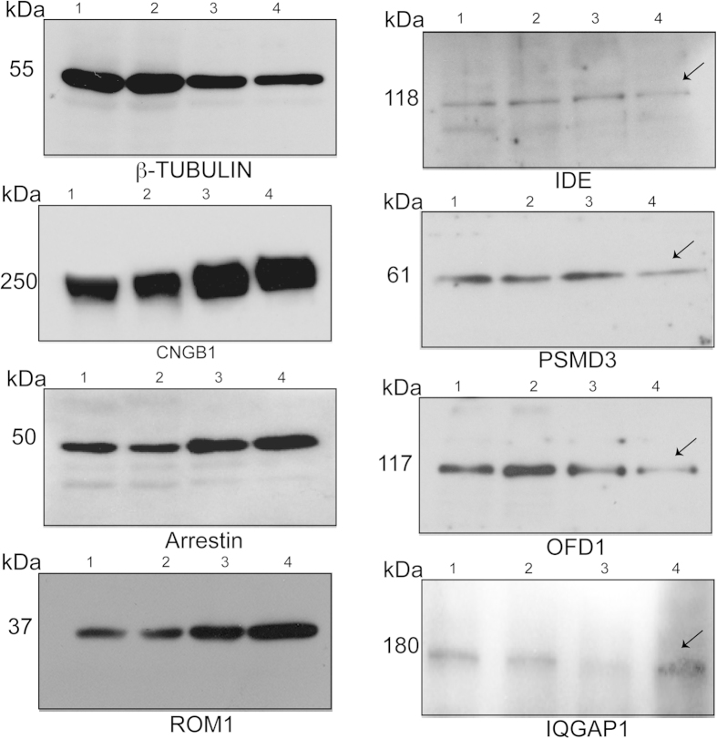
Figure 3. Validation of selected proteins identified by MS/MS analysis.
PSC from wild type (WT) and Rpgrko mouse retina were analyzed by SDS-PAGE and immunoblotting using indicated antibodies. Lanes 1 and 2: total retina lysate from WT and Rpgrko mice, respectively; Lanes 3 and 4: PSC from WT and Rpgrko mice, respectively. Arrows indicate altered proteins in the Rpgrko PSC. Molecular weight markers are shown in kilo Daltons (kDa).

