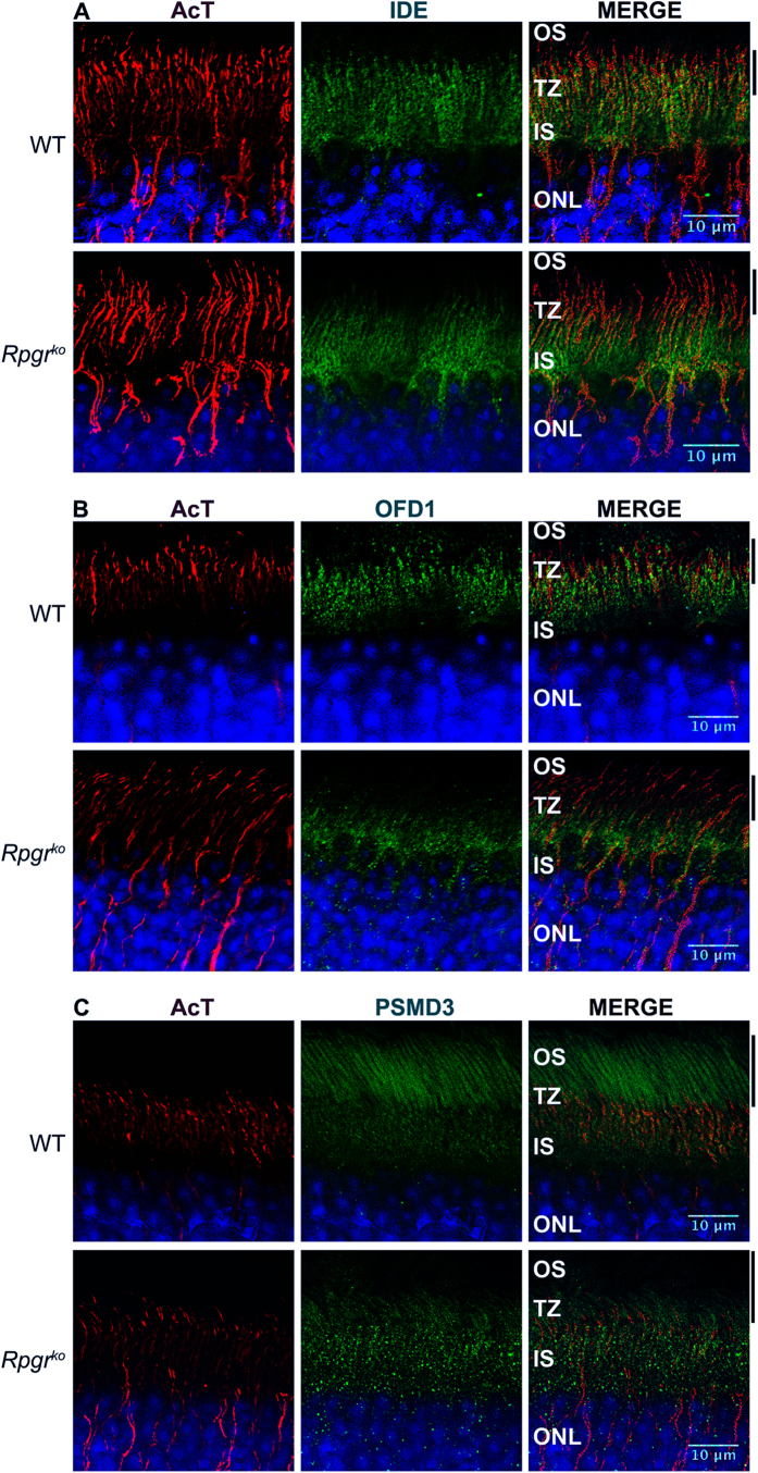
Figure 4. Localization of PSC proteins in mouse retina.
Wild type (WT) and Rpgrko retinal sections were stained with indicated antibodies. Nuclei (blue) are stained with Hoechst. OS: outer segment; TZ: transition zone; IS: inner segment; ONL: outer nuclear layer; AcT: acetylated α-tubulin. Scale bar: 10 μm. Solid lines indicate the area that is stained by the respective antibody (green) in WT but not in Rpgrko retina.

