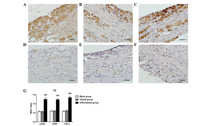
Figure 2.
Representative images of p-ERK, CGRP and COX-2 immunoreactivity in the TG 30 min after (A–C) NTG or (D–F) vehicle infusion by immunohistochemistry and (G) analysis of the MOD of p-ERK, CGRP and COX-2 expression in the TG. An increase in (A) p-ERK, (B) CGRP and (C) COX-2 expression was observed in the TG following NTG infusion and the MOD value for NTG-treated rats was significantly higher than in the vehicle and blank groups. (###P<0.001, compared with the vehicle and blank groups; n=6 in each group; error bars indicate standard deviation; scale bar=100 µm). MOD, mean optical density; p-ERK, phosphorylated extracellular signal-regulated kinase; CGRP, calcitonin gene-related peptide; COX-2, cyclooxygenase-2; TG, trigeminal ganglion; NTG, nitroglycerin.

