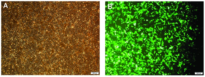
Figure 2.

Representative microscopy images of CNE-2 cells following PARP-1-silencing by lentivirus-delivered small-hairpin RNA transfection. (A) Phase contrast image of transfected CNE-2 cells. (B) Fluorescence microscopy image of green fluorescent protein expression of cells. Images were captured of cells in an identical field of vision (scale bar, 200 µm). PARP-1, poly-(adenosine diphosphate-ribose) polymerase-1.
