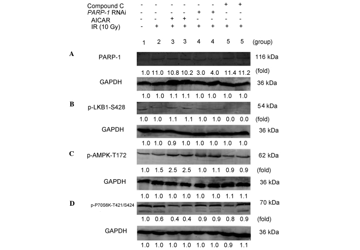
Figure 3.
PARP-1 promotes autophagy through the AMPK/mammalian target of rapamycin pathway in CNE-2 cells following IR. CNE-2 cells in each group were treated as follows: 1, untreated control group); 2, 10 Gy IR; 3, pretreatment with AICAR (2.0 mM) for 2 h + 10 Gy IR; 4, PARP-1 RNAi using lentivirus-delivered small-hairpin RNA transfection + 10 Gy IR; 5, pretreatment with Compound C (10 µM) for 2 h + 10 Gy IR. (A) After 48 h, CNE-2 cells were collected and subjected to western blot analysis for the detection of PARP-1. After 30 min, CNE-2 cells were collected and subjected to western blot for detection of (B) p-LKB1-S428, (C) p-AMPK-Thr172 and (D) p-P70S6K-T421/S424. GAPDH was used as the loading control. Protein expression levels of PARP-1, p-LKB1-Ser428, p-AMPK-Thr172 and p-P70S6K-T421/S424 were then quantified and the mean fold change relative to GAPDH for three independent experiments. PARP-1, poly-(adenosine diphosphate-ribose) polymerase-1; AMPK, adenosine monophosphate-activated protein kinase; IR, ionizing radiation; AICAR, 5-amino-1-β-D-ribofuranosyl-1H-imidazole-4-carboxamide; RNAi, RNA interference; Compound C, 6-[4-(2-Piperidin-1-yl-ethoxy)-phenyl)]-3-pyridin-4-yl-pyrrazolo [1,5-a]-pyrimidine; LKB1, liver kinase B1, p-, phosphorylated.

