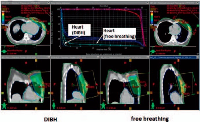
Fig. 1.
Dose distribution in radiotherapy of the left breast during free breathing (left) and DIBH technique (right). The 3-dimensional dose distributions on corresponding CT sections (same height in the chest) show that the contours of the heart are different and the distance between the heart and the radiation field is greater for inspiration. This leads to a lower radiation dose to the heart, evident in the dose-volume histogram (top center). The median heart dose is almost identical, but the maximum dose is significantly reduced by the DIBH technique.

