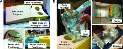
FIG. 1.

Apparatuses for phantom experiments evaluating prone and supine PET/CT. (A) First, a rigid phantom study characterized effects of prone support device on lesion detection. Top, prototype prone positioner for PET and MR imaging of breast consists of a soft foam support ramp, a rigid foam upper torso support with breast cutouts covered with a soft foam pad. Bottom left, breast phantom prone in the positioning device, in the CT field of view. Bottom right, phantom without the positioning device. The prone positioning device is seen on the right side for comparison of relative phantom height with and without the device; the prone positioner was removed from the scanner bed for the without-positioner scan. (B) Second, a nonrigid phantom study evaluated position-dependent differences in attenuation between prone and supine images. Left, positioning of body phantom with affixed intravenous (I.V.) saline bags (mock breasts) containing 18F-FDG hanging pendant prior to placement in the scanner. Top right, for prone scans, I.V. bags were positioned inside the prone positioner cutouts and the body phantom was placed on top of the torso support. Bottom right, for supine scans, the positioner was removed and I.V. bags were taped adjacent to the anterior wall of the body phantom.
