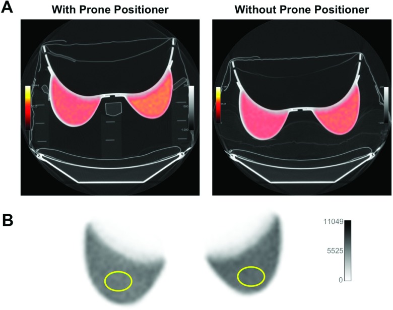
FIG. 2.
Characterization of prone positioner with rigid breast phantom. (A) PET/CT of breast phantom with (left) and without (right) the prone positioner. PET images were fused with CT scans set to a pulmonary gray scale level for visualization of low-density materials of the positioner and pillow support in the scan without positioner. PET images were scaled to compensate for the activity decay between scans (22 min). (B) Elliptical ROIs (11.8 cm2) were drawn over each side of the phantom to measure activity concentration. Values in Table I represent the average of 15 ROI means (from one side of the phantom) over 15 image slices.

