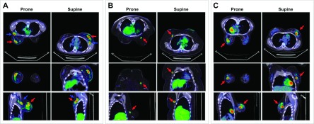
FIG. 7.

Fused PET/CT images comparing prone and supine positions. (A) 36-yr old patient (P21) with Stage IIIA, intermediate-grade, node-positive LABC, in whom the prone-supine SUVpeak percentage difference (P–S%Δ), after correcting for uptake time differences (ti = 60 min), closely approximated the corrected group mean P–S%Δ: P21, 11.4% (0.92 SUV) versus group 11.4% (0.82 SUV). (B) 46-yr old patient (P32) with ER-and HER2-negative, high-grade LABC in whom P–S%Δ was the largest in the corrected group: 34.7% (2.01 SUV). (C) 32-yr old patient with ER-/PR-/HER2-positive, high-grade LABC in whom P–S%Δ was the smallest in the corrected group: 0.24% (0.018 SUV). Relative to supine position, prone FDG PET/CT with the positioner improved the separation of primary tumor from axilla and chest wall (arrows).
