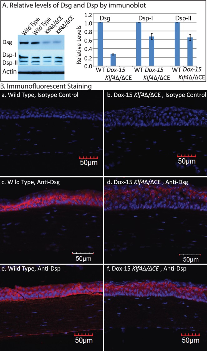
Figure 7.

Decreased expression of desmosomal components in the Dox-15 Klf4Δ/ΔCE corneas. Immunoblots (A) revealed decreased amount of Dsg, Dsp-I, and Dsp-II in the Klf4Δ/ΔCE corneas after 15 days of doxycycline treatment. Densitometry with actin as loading control quantified the amounts of Dsg, Dsp-I, and Dsp-II within the Dox-15 Klf4Δ/ΔCE corneas to be approximately 30%, 70%, and 70% of that in the WT, respectively. Consistent with this, immunofluorescent staining (B) detected robust expression of Dsg, Dsp-I, and Dsp-II in the WT (red; [B-c, B-e], respectively), which was decreased in the Dox-15 Klf4Δ/ΔCE (B-d, B-f) corneal epithelium. No background staining was observed in isotype antibody control (B-a, B-b).
