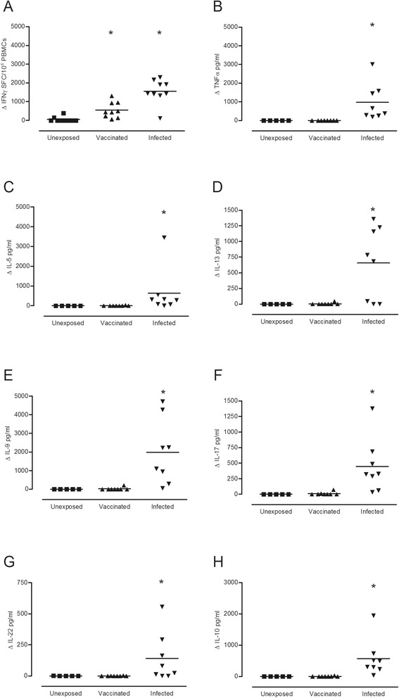
Figure 1.

Differential cytokine responses to anthrax LF domain IV following cutaneous infection or AVP vaccination. Cells from individuals exposed to LF as a result of (▼) natural cutaneous infection (n = 8–9), or (▲) AVP vaccination (n = 8–10) and (■) unexposed healthy controls (n = 5-10) were stimulated with 25 μg/ml of LF domain IV in vitro, and the cytokine profile of the supernatants assessed by either ELIspot, Luminex or ELISA. ELIspot results (A) are expressed as the mean ΔSpot Forming Cells (SFC)/106 PBMCs (stimulated – unstimulated background level), while the ELISA and Luminex values are given as the mean Δpg/ml detected for (B) TNFα, (C) IL-5, (D) IL-13, (E) IL-9, (F) IL-17, (G) IL-22 and (H) IL-10. * denotes a significantly greater cytokine production in comparison to the unexposed controls (p ≤ 0.05), as determined by Kruskal Wallis with Dunns multiple comparison test performed using GraphPad Prism version 5.01 for Windows, GraphPad Software, La Jolla California USA.
