Figure 4.
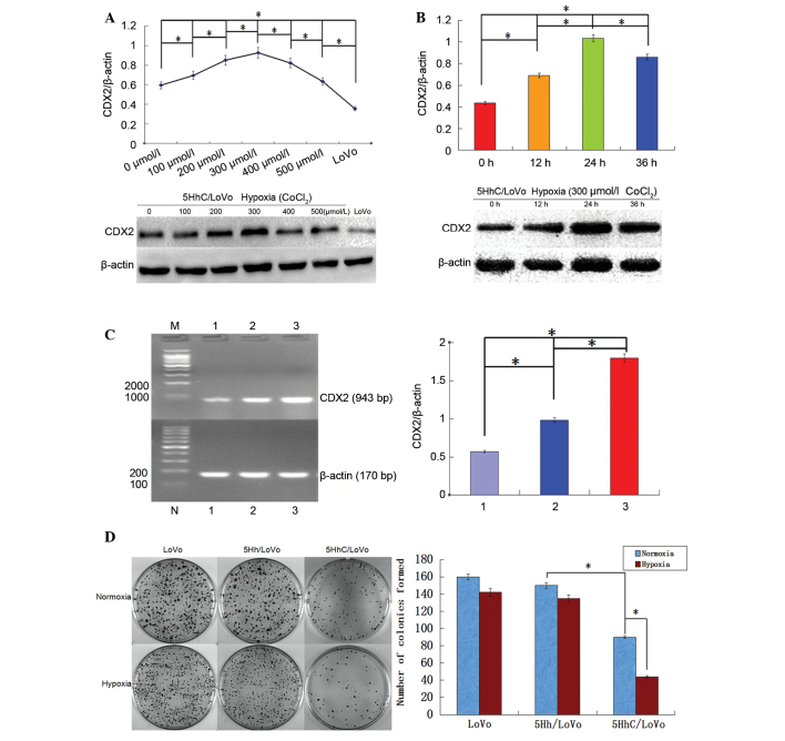
Expression levels of CDX2 in 5HhC LoVo cells and the effect of CDX2 overexpression on LoVo cell proliferation. (A) Western blot analysis of CDX2 expression. The 5HhC/LoVo cells and LoVo cells were cultured under normoxic or hypoxic conditions (100–500 µmol/l CoCl2) for 24 h. (B) Western blot analysis of CDX2 expression. The 5HhC/LoVo cells were cultured under hypoxic conditions (300 µmol/l CoCl2) for 0, 12, 24 or 36 h. (C) Reverse transcription-polymerase chain reaction analysis of CDX2 mRNA expression. The 5HhC/LoVo cells were cultured under a normoxic or hypoxic conditions (300 µmol/l CoCl2) for 24 h. M, DNA marker; 1, LoVo cells; 2, 5HhC/LoVo cells under normoxic conditions; 3, 5HhC/LoVo cells under hypoxic conditions. (D) Clone formation of LoVo, 5Hh/LoVo or 5HhC/LoVo cells. Each group had a hypoxic control (200 µmol/l CoCl2; red bars). All data presented are representative images of each group of cells from three separate experiments. The results are presented as the mean ± standard deviation (*P<0.05). CDX2, caudal-related homeobox; 5HhC, pLVX-5HRE-hTERTp-CDX2-3FLAG; 5Hh, pLVX-5HRE-hTERTp-EGFP-3FLAG; bp, base pairs.

