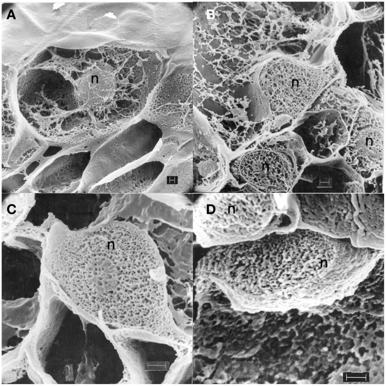
Figure 2.
Scanning electron micrographs (Hadwiger and Adams, 1978) showing cross-sections of freeze-fractured, freeze dried pea pod epidermal surface cells from endocarp tissue: Untreated (A), treated with water 1 h (B), inoculated with macroconidia of Fusarium solani f. sp. phaseoli 1 h (C), and 6 h (D). Pea nuclei, n; fungus, f; EP, Epidermis surface. Bar = 1.0 μ.

