Figure 5.
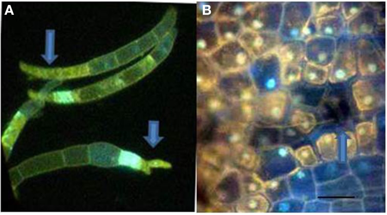
F. solani f.sp. phaseoli (Fsph) spores DAPI stained to detect fungal nuclear and plant cell DNA positive material. (A) Fsph spores cultured in shake culture 3.5 h followed by contact with pea endocarp tissue for 2 h, viewed under UV light. Arrows point to DAPI stained tips of the spores where nuclear destruction is evident. Some spore cell compartments were devoid of detectable nuclei. (B) Photo of epidermal surface of a Fsph lesion encompassing both non-flourescing plant cells and a non-fluorescing, non-visible spore 20 h pi. The fluorescing nuclei in cells surrounding the spore indicate different states of the plant nuclear staining depending on their location away from the lesion. Bar = 30μ.

