
Figure 2.
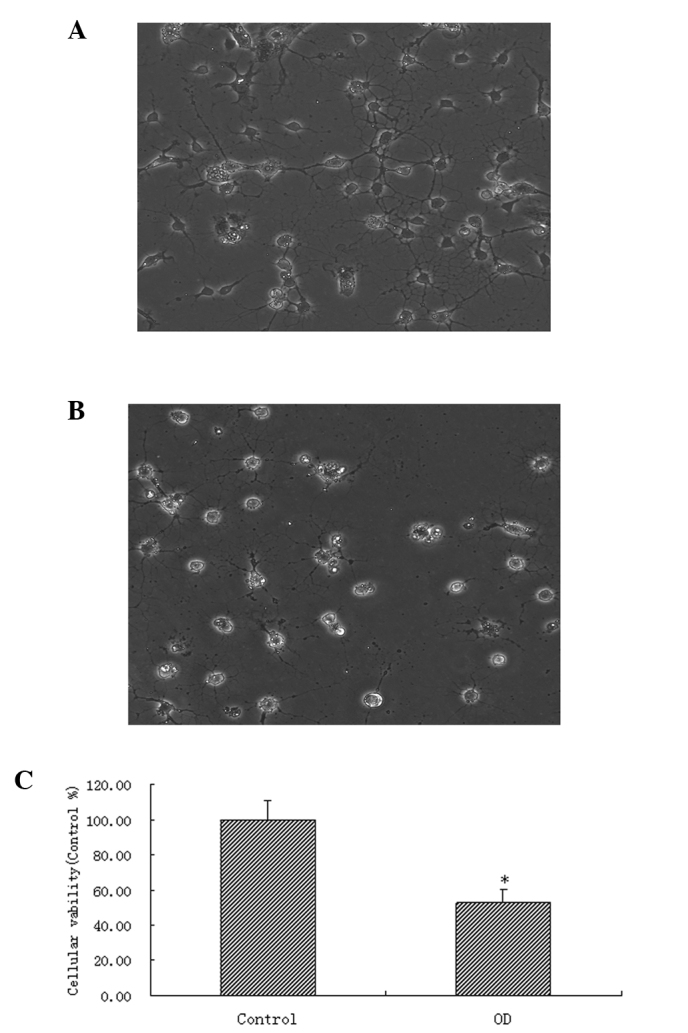
Establishment of a model of OGD. (A) Spinal motor neurons thawed after 72 h, with normal cell bodies and numerous continuous projections (×200). (B) Cells following OGD injury, with various degrees of cell body swelling and breakage of projections (×200). (C) MTT assay of cell viability in each group.*P<0.05, compared with the control group. OGD, oxygen and glucose deprivation.