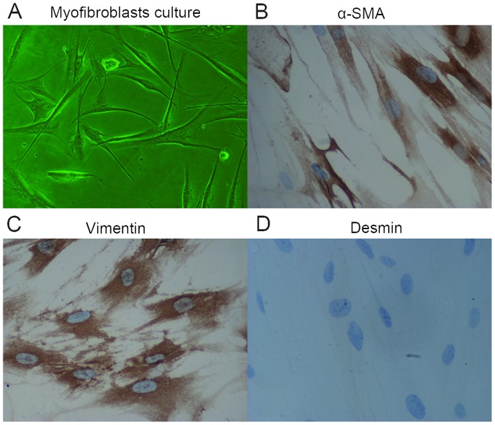
Figure 4.
Characterization of isolated subepithelial myofibroblasts. (A) Images of isolated myofibroblasts (magnification, x100). Immunostaining with monoclonal antibodies. (B) Positive staining for α-SMA; (C) positive staining for vimentin; and (D) negative staining for desmin. Magnification, x400. α-SMA, α-smooth muscle actin.

