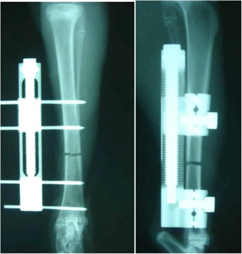
Fig. 1.

Schematic representation of the callus distraction model. The Orthofix M-100 was fixed to the tibia with four half pins of 2.0 mm. The middle two pins were set at 20 mm apart, and at the center of these, a drill hole osteotomy was performed using a 1.0-mm drill. In order to fit the fragments, an extender is turned in the shortening direction, and the bone fragments were brought into contact with each other
