Fig. 5.

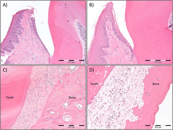
Morphologic changes of gingiva and periodontal membrane six weeks after 5 × 15 Gy radiotherapy. H&E stained sections of the gingiva of non-irradiated control rats (a) and 5 × 15 Gy irradiated rats (b). The histologic examination demonstrates epithelial transformation from slightly parakeratinized in controls to hyperkeratinized and hyperplasic in irradiated rats. This is also evident in the epithelium closest to the tooth. In addition the connective tissue is more fibrous and contains fewer cells after irradiation. Scale bar indicates 200 μm. (c) H&E stained sections of the periodontal membrane of non-irradiated control rats. The periodontal membrane is richly vascularized with fibers organized for proper attachment of tooth to bone. Scale bar indicates 200 μm. (d) H&E stained sections of the periodontal membrane of 5 x15 Gy irradiated rats. The irradiated periodontal membrane demonstrates fibrous connective tissue with few cells, unorganized fibers and infiltration of mononuclear inflammatory cells. Scale bar indicates 200 μm
