
Fig. 8.
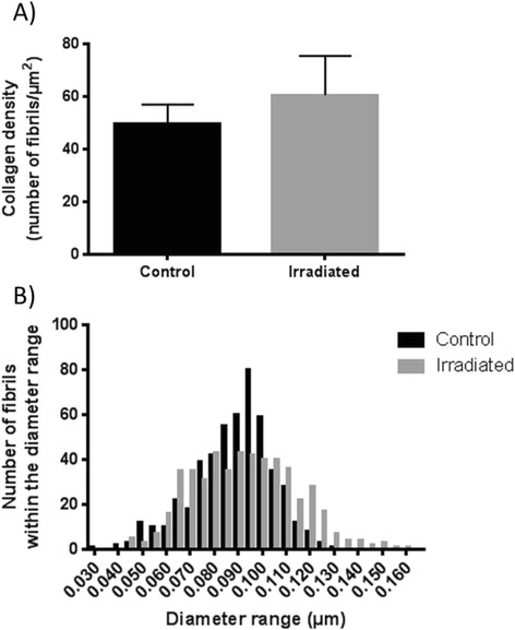
Collagen density and diameter six weeks after 5 × 15 Gy radiotherapy. (a) The average fibril density in control (n = 5) and irradiated (n = 5) skin samples from the outer part of the radiation field presented as mean ± SD. (b) The frequency distribution of collagen fibril diameter demonstrates a minor displacement towards larger fibril diameter in the skin after radiotherapy (n = 5) compared to control animals (n = 5)