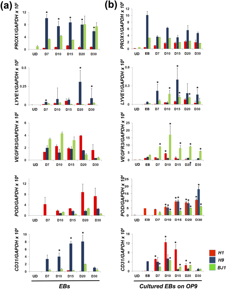
Figure 1. Differentiation of hESCs (H1 and H9) and hiPSCs (BJ1) into the LEC lineage.
(a) qRT-PCR analyses of in vitro differentiated H1, H9 and BJ1 cells through EB formation. N = 9 per group. *P < 0.05 vs. H1. (b) qRT-PCR analyses of in vitro differentiated H1, H9 and BJ1 cells on OP9 cells with lymphangiogenic cytokines. EBs differentiated for 7 days in suspension culture were replated on OP9 cells, further cultured for an additional 30 days with VEGF-A, -C and EGF, and subjected to qRT-PCR. N = 9 per group. *P < 0.05 vs. EB. UD: Undifferentiated hPSCs, POD: PODOPLANIN.

