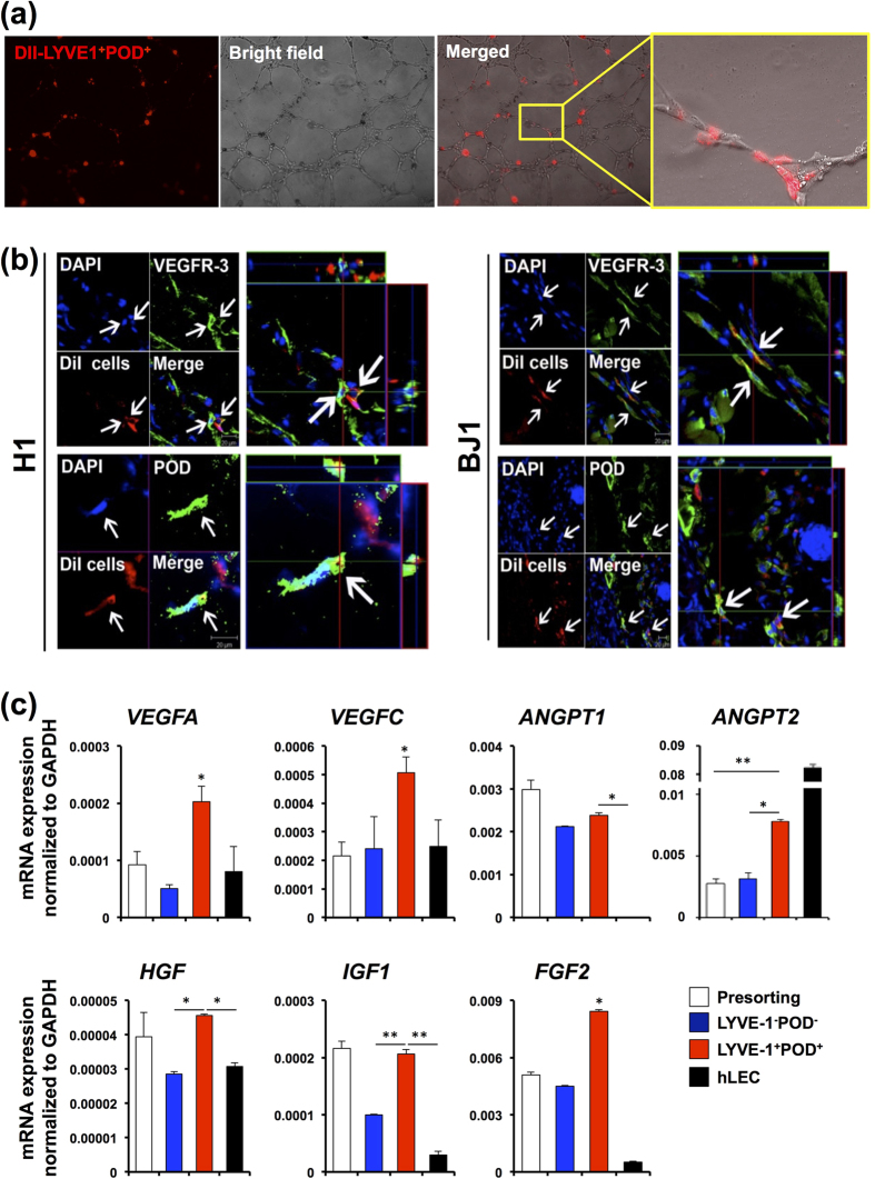
Figure 4. Contribution of LYVE-1+PODOPLANIN+cells derived from hESCs and hiPSCs to new lymphatic vessel formation.
(a) In vitro tube formation assay. DiI-labeled LYVE-1+PODOPLANIN+cells (red) derived from hiESC (H1) cocultured with hLECs onto Matrigel for 12 hours, showing their incorporation into vascular structures consisting of hLECs. (b) DiI-labeled LYVE-1+PODOPLANIN+cells which were differentiated from H1 and BJ1 cells were injected into mice in an ear wound model. Two weeks later, tissues were harvested and subjected to immunohistochemistry with VEGFR3, PODOPLANIN, and LYVE-1 antibodies on frozen tissue sections. Confocal microscopic imaging revealed incorporation of injected cells (red) into lymphatic vascular structures and expression of PODOPLANIN and VEGFR3. (c) mRNA expression of the indicated lymphangiogenic genes in pre-sorted cells at day 14, LYVE1+PODOPLANIN+cells (BJ1-derived), LYVE1−PODOPLANIN−cells (BJ1-derived), and hLECs measured by qRT-PCR. Data were presented as relative mRNA expression to GAPDH. N = 3 to 4 per group. *P < 0.05. **P < 0.01.

