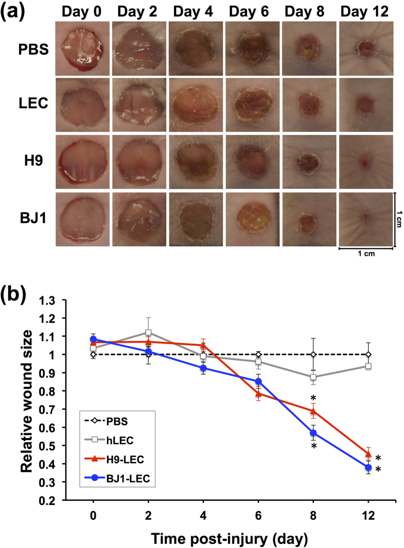
Figure 5. hPSC-derived LECs improve wound healing and increase lymphatic vessel formation.
FACS-sorted LYVE-1+PODOPLANIN+cells derived from hPSCs (H9 and BJ1) were injected into skin wound on the backs of nude mice. PBS and hLECs were used as controls. (a) Changes of the wound areas at indicated days after injection. (b) Quantitative analysis of wound areas. Y axis represents percent change of wound areas over original wound areas. Note that LYVE-1+PODOPLANIN+cells derived from both H9 and BJ1 significantly promoted wound closure compared to the PBS- or hLEC-control. Two independent experiments were performed. N = 5 to 9 per group. *P < 0.05, **P < 0.01.

