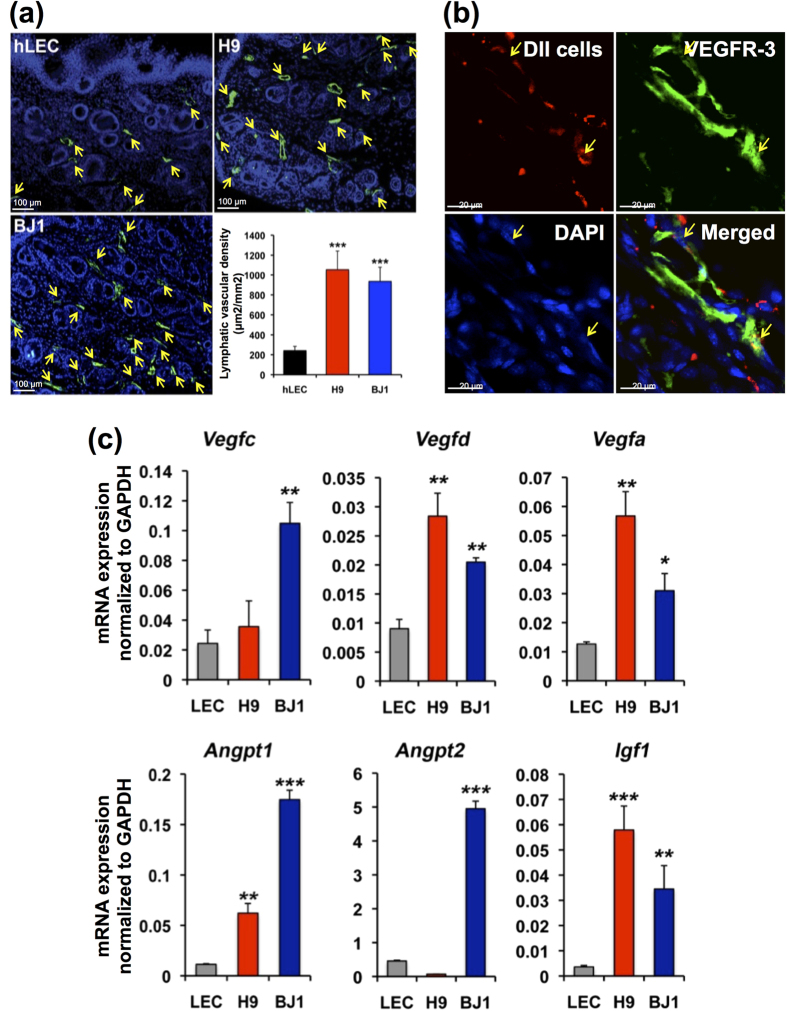
Figure 6. Lymphvasculogenesis and lymphangiogenesis induced by hPSC-LECs in wound healing.
(a) LYVE-1+PODOPLANIN+cells derived from hPSCs (H9 and BJ1) were injected into skin wound on the backs of nude mice. Lymphatic vascular density determined at day 14 after cell injection. Lymphatic vessels indicated by arrows (LYVE-1+) were significantly increased in mice injected with hPSC-derived LYVE-1+PODOPLANIN+cells compared to the hLEC-control. ***P < 0.001. Polyclonal rabbit anti-LYVE-1 antibody was used for this staining. (b) LYVE-1+PODOPLANIN+cells labeled with CM-Dil were injected into the wounded skin in mice. Confocal microscopic imaging after staining tissues harvested at 2 weeks for VEGFR3 (green) showed incorporation of the injected cells (red) into lymphatic vessels. Arrows indicate co-localization of VEGFR3+lymphatic vessels with the injected cells. **P < 0.01. (c) Skin tissues from mice injected with hLEC, H9-LEC, or BJ1-LEC were subjected to qRT-PCR analyses. Graphs from 3 independent experiments are shown, *P < 0.05, **P < 0.01, ***P < 0.001, N = 3 per group.

