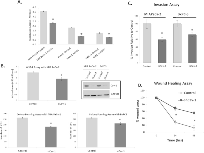
Figure 2. Cav-1 is essential for proliferation, invasion, and migration of pancreatic cancer cells.
A. Pre-treatment of MIAPaCa-2, Panc-1 and BxPC3 cell lines with methyl-β-cyclodextran (MβCD) at 5 mM for 1 hour results in reductions in proliferation as measured by WST-1 assay (48 hrs). Absorbance is plotted on the y-axis where readings at 450 nm are subtracted from background reading at 600 nm, (*p < 0.05; n = 3). B. WST-1 assay comparing treatment with scrambled siRNA (“control”) and Cav-1 siRNA (“siCav-1”) in MIAPaCa-2 cells (top panel). Colony forming assays after treatment with scrambled control siRNA or Cav-1 siRNA on MIAPaCa-2 and BxPC3 cell lines (p < 0.05, n = 3, bottom panels). Western blot (top left) confirms appropriate Cav-1 depletion with Cav-1 siRNA in both cell lines. Virtually identical results were observed with a completely distinct set of pooled siRNA to Cav-1 (data not shown). C. Cells were seeded in 1% FBS-containing medium into the tops of Transwell chambers coated with Matrigel and incubated for 24 hours before being washed and fixed. Chemoattractant in the bottom well was 10% FBS. Columns represent percentage of invasion relative to control siRNA for different cell lines pre-treated with scrambled siRNA (control) and siCav-1 siRNA pools for 48 hours (left panel, p < 0.05, n = 4). D. Scratch (wound-healing) assay to measure migration, showing the percentage wound area plotted against time for scrambled siRNA (control)and siCav-1 treated MIA PaCa-2 cells (right panel). Note that control cells show almost complete closure of the “wound” compared to Cav-1 knockdown cells by 48 hours, suggesting a migration defect in Cav-1 depleted cells (*p < 0.05; n = 3).

