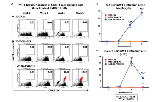
Figure 4.

mWT1-specific cytotoxic T-lymphocyte induction by caTLR4-PMDC11 cells. Normal PB-CD8+ T cells were cultured with mWT1 peptide-pulsed PMDC11, LPS-stimulated PMDC11 or doxycycline-exposed caTLR4-PMDC11 cells for five weeks. The co-cultured cells were double stained with the FITC-conjugated CD8 antibody and PE-conjugated mWT1 tetramer. (A) mWT1 tetramer analysis of CD8+ T cells cultured with PMDC11, LPS-stimulated PMDC11 or doxycycline-exposed caTLR4-PMDC11 cells. Values in the dot plots represent the percentages of mWT1 tetramer+/CD8+ T cells (marked in red) among the cells in the lymphocyte gate of forward scatter/side scatter dot plots. Dot plots are representative of three experiments with similar results. (B) Percentages of mWT1 tetramer+/CD8+ T cells in the lymphocyte gate of the forward scatter/side scatter dot plots. (C) Total number (×104) of CD8+ mWT1 tetramer+ cells in a well, which was calculated by the formula [(percentage of mWT1 tetramer+/CD8+ T cells among lymphocytes) × (total number of lymphocytes in culture well)]. LPS, lipopolysaccharide; FITC, fluorescein isothiocyanate; PE, phycoerythrin.
