
Figure 6.
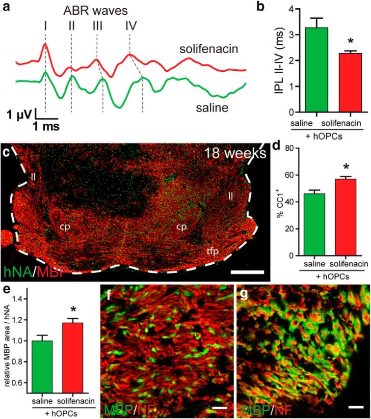
Solifenacin improves functional recovery mediated by transplanted hOPCs. To determine whether solifenacin treatment improves the function of myelin produced by transplanted hOPCs, we transplanted hOPCs into the midbrain of shiverer/rag2 day 1–3 pups and assessed auditory brainstem response at 15 weeks after daily subcutaneous injection with either saline or 10 mg/kg solifenacin. a, Auditory evoked potential waveform from saline- and solifenacin-treated hOPC-transplanted animals indicating the relative timing of positive vertex waves I-IV. b, IPL between peaks II and IV was measured (mean ± SEM, n = 3–5). Solifenacin treatment significantly reduced IPL II-IV. *p < 0.05, t test. At 19 weeks, animals were killed to determine the distribution of human cells (hNA, green) and myelin (MBP, red). c, Myelin was found throughout the ventral midbrain structures, including cerebral peduncle (cp), lateral lemniscus (ll), and transverse fibers pons (tfp). The example coronal section shown from a solifenacin-treated animal corresponds to plate 65 in Franklin and Paxinos (2008). d, Proportion of human cells differentiating as CC1+ oligodendrocytes in the cerebral peduncle. e, Relative contribution of human cells to percentage area of MBP staining in the cerebral peduncle. f, g, Confocal images of myelinated fibers in the cerebral peduncle of saline-treated (f) and solifenacin-treated (g) animals at 19 weeks (neurofilament, red; MBP, green). Scale bars: 500 μm (c), 10 μm (f–g).