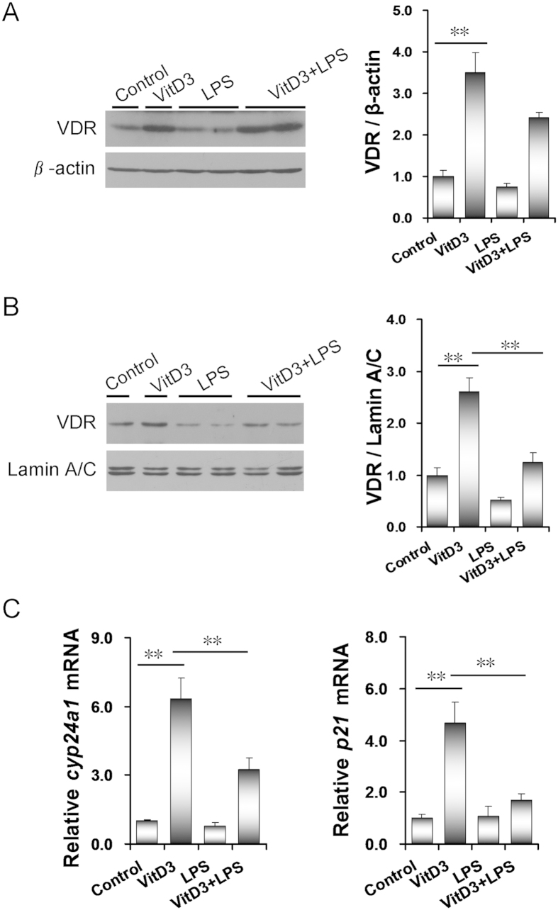
Figure 6. LPS inhibits VitD3-activated placental VDR signaling.
All pregnant mice were i.p. injected with LPS (100 μg/kg) on GD15. In VitD3 + LPS group, pregnant mice were pretreated with vitamin D3 (25 μg/kg) 24 h and 1 h before LPS injection. Placentas were collected 2 h after LPS injection. (A) Placental VDR was detected using immunoblots. (B) Nuclear VDR was detected using immunoblots. All experiments were duplicated for four times. A representative gel for VDR (upper panel) and lamin A/C (lower panel) was shown. All data were expressed as means ± S.E.M. (n = 6–8). (C) Placental cyp24a1 and p21 mRNA was measured using real-time RT-PCR. All data were expressed as means ± S.E.M. (n = 8). ** P < 0.01.

