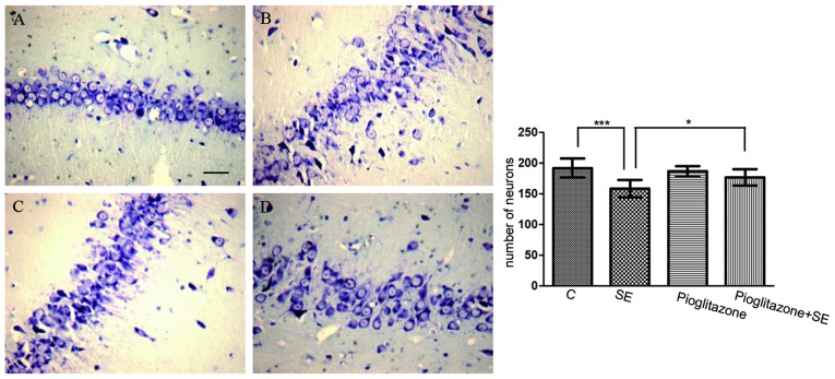
Figure 2.
Neuronal loss in the hippocampal CA3 area following SE. Neuronal damage was assessed histologically using Nissl staining. Compared with the (A) control group, more dark colored, disorganized and blurred Nissl bodies were found in the (B) vehicle-treated SE group on the third day following SE, (C) slightly blurred Nissl bodies were observed in the pioglitazone group and the number of Nissl bodies decreased in the (D) pioglitazone-treated SE group. Scale bar=20 µm. Data are expressed as the mean ± standard deviation. *P<0.05 and ***P<0.001. C, control; SE, status epilepticus.

