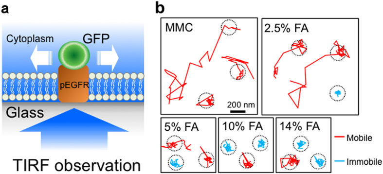
Figure 1. Single-molecule measurement of a membrane protein in FA-fixed MEFs.

(a) Schematic of the method for single-molecule measurement. EGFR-GFP molecules expressed in MEFs were observed at a single-molecule resolution by TIRF microscopy after MMC treatment or FA fixation. (b) Examples of single-molecule movements of EGFR-GFP. The molecular movement was traced at a temporal resolution of 30.5 msec for 6 s (200 frames). In this measurement, molecules that expanded in the >200-nm range and stayed in the <200-nm range were regarded as mobile and immobile molecules, respectively. Dotted circles represent the 200-nm range.
