
Figure 7. Subcellular localization of KV7.1 and mutants.
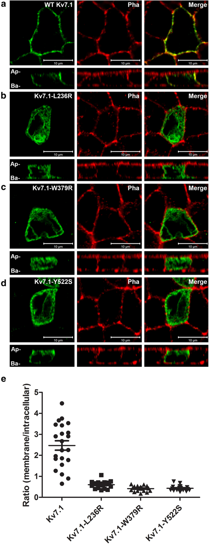
Horizontal and vertical confocal images of polarized MDCK cells transiently expressing the KV7.1–WT or MUT as indicated and labeled with antibodies against KV7.1 (left panels) and Phalloidin (middle panels). Merged pictures are shown in right panels. Ap; apical; Ba, basolateral. Representative pictures from three independent experiments are shown. Quantification of ratio between channels in the membrane versus channels trapped in intracellular compartments (most likely ER) for WT and mutants (p < 0.0001, n = 18−22 cells each situation).