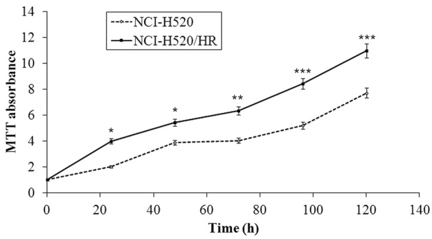
Figure 3.

Cell proliferation assays of NCI-H520 and NCI-H520/R cells. The cells were cultured in 96-well plates for 0, 24, 48, 72, 96, and 120 h. The proliferation of the cells was determined using an MTT assay. The data are expressed as the mean ± standard deviation of three experiments. Statistically significant differences were obtained (*P<0.05;**P<0.01; ***P<0.001; n=6). NCI-H520/R, radioresistant subclone.
