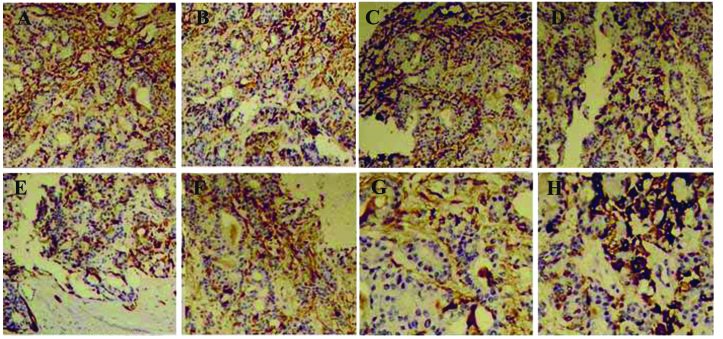
Figure 5.
Histochemical analysis of TNF-α expression in the periodontal tissue in the diabetes group. There was positive expression of TNF-α in (A) the gingival epithelial layer, (B) the granular layer and stratum spinosum, (C) fibroblasts, (D) vascular endothelial cells, (E) osteoblasts, (F) osteoclasts, (G) the cytoplasm of bone marrow stromal cells and (H) the gingival epithelial basal layer. Magnification, ×400. TNF, tumor necrosis factor.

