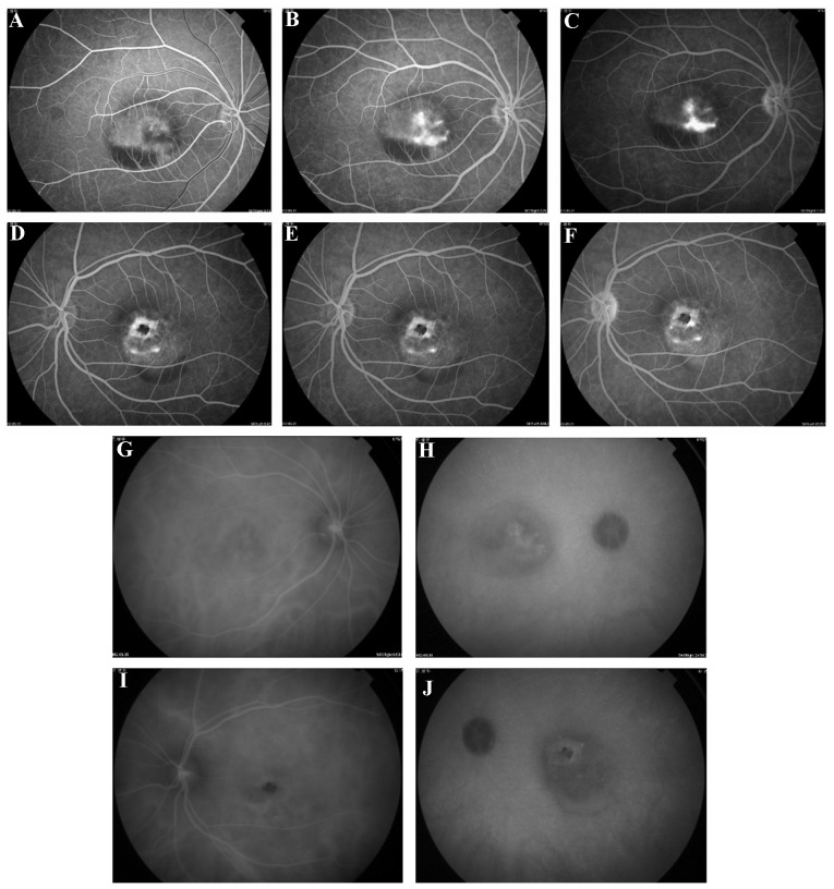
Figure 2.
Fundus photography, FFA imaging and ICG were performed using a retina angiograph. (A) FFA revealed significant early hyperfluorescence (B and C) that increased in intensity at the late stage of the angiographic sequence (D–F) and was associated with moderate leakage in the right eye and macular lesions that simulated a pattern of dystrophy in the left eye. (G and I) ICG detected hypofluorescence in the early stages of the angiographic sequence in the left and right eyes and subsequently detected (H and J) little abnormal hyperfluorescence at the late stages (magnification, ×50). ICG, indocyanine green chorioangiography; FFA, fundus fluorescein angiography.

