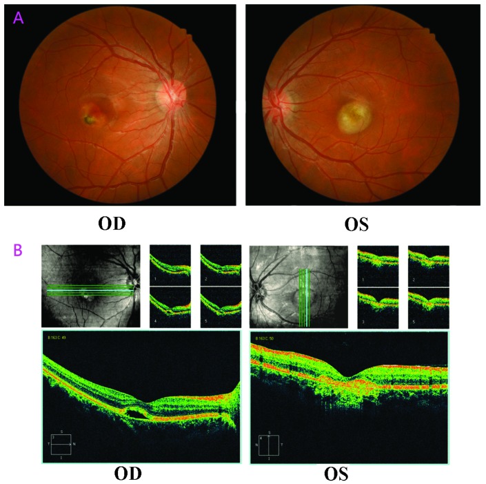
Figure 4.
(A) Examination of the fundus of Case 2. Vitelliruptive lesions with a scrambled egg-like appearance were observed, in addition to dispersion of the vitelliform material with signs of atrophy in the right eye. (B) Spectral domain optical coherence tomography scans demonstrated clear abnormalities, including the absence of the foveal pit, serous retinal detachment, cystoid macular edema and interruption of the outer limiting membrane. The right eye exhibited marked yellow-white subretinal scarring with pigmented borders, surrounded by serous retinal detachment. In addition, the left eye exhibited an atrophic lesion (magnification, ×50). OD, oculus dexter; OS, oculus sinister.

