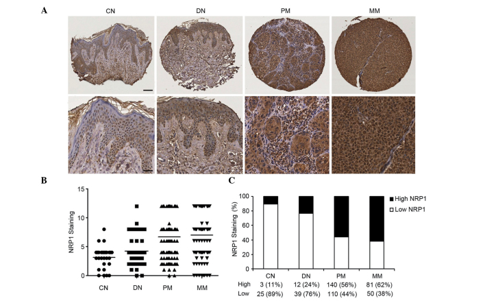
Figure 2.
Increased NRP1 expression is correlated with melanoma progression. (A) Representative images of CN and DN, with low NRP1 expression, and PM and MM, with high NRP1 expression (upper panel, scale bar 40 µm; lower panel, scale bar 20 µm). (B) Kruskal-Wallis test for differences in NRP1 staining among CN, DN, PM and MM. The mean is depicted as a horizontal line in each group (n=460, P<0.0001). (C) NRP1 expression was increased from CN to DN, PM and MM (n=460, P=3.6×10⁻⁹, χ2 test). Magnification, ×100 (upper panel), ×200 (lower panel). NRP1, neuropilin 1; CN, common nevi; DN, dysplastic nevi; PM, primary melanoma; MM, metastatic melanoma.

