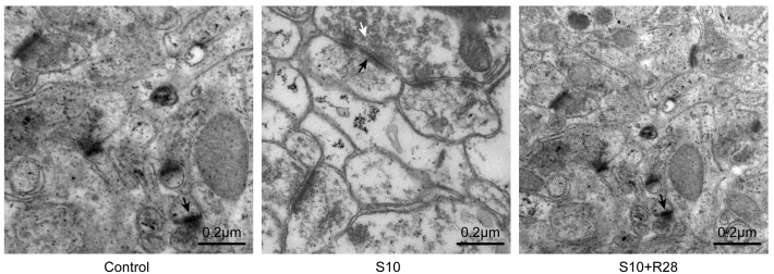
Figure 3.

Ultrastructural alterations in synapses of hippocampal Cornu Ammonis 1 neurons in salicylate-treated animals. Representative images are shown at magnification, ×30,000. Transmission electron micrographs show that animals in the S10 group had a greater number of presynaptic vesicles (white arrows), thicker postsynaptic densities (black arrows), and greater synaptic interface curvature, as well as fewer microtubules and neurofilaments (arrowheads), than saline-injected control animals. S10, chronic salicylate treatment group; R#, number of recovery days.
