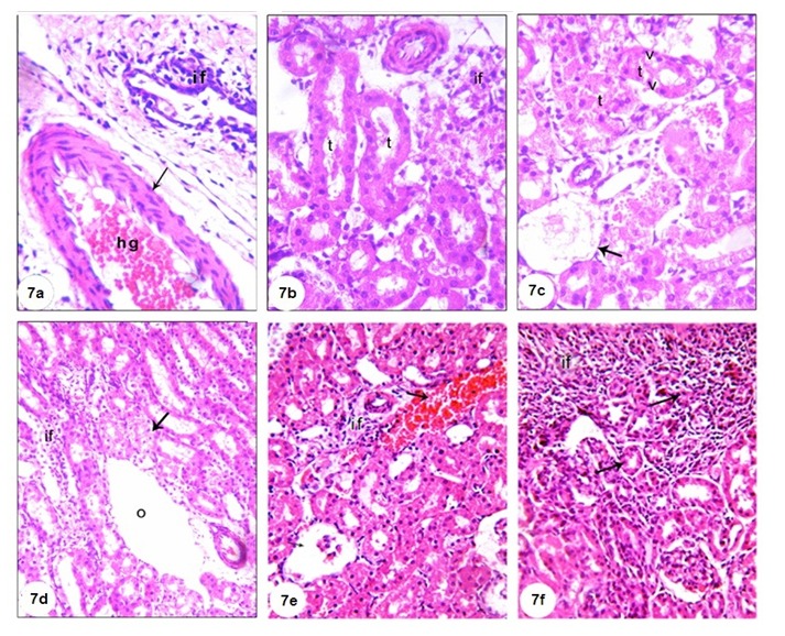
Figure 7. A photomicrograph of H&E stained kidney section of GM-administered rats (Group 2) showing dilated hypermic portal vein (hg), inflammatory cells infiltration (if) (7a), damaged and dilated tubule (t) with if (7b), complete atrophy of some glomeruli (arrow), desquamation of tubular epithelium with cytoplasmic vaculations (v) (7c), odema (o) with a number of inflammatory cells, proximal tubular necrosis (arrow) (7d), and interstitial haemorrhage (arrow) accompanied with proximal tubular necrosis and shrinkage of glomerular capillary (7e and f), (X400).

