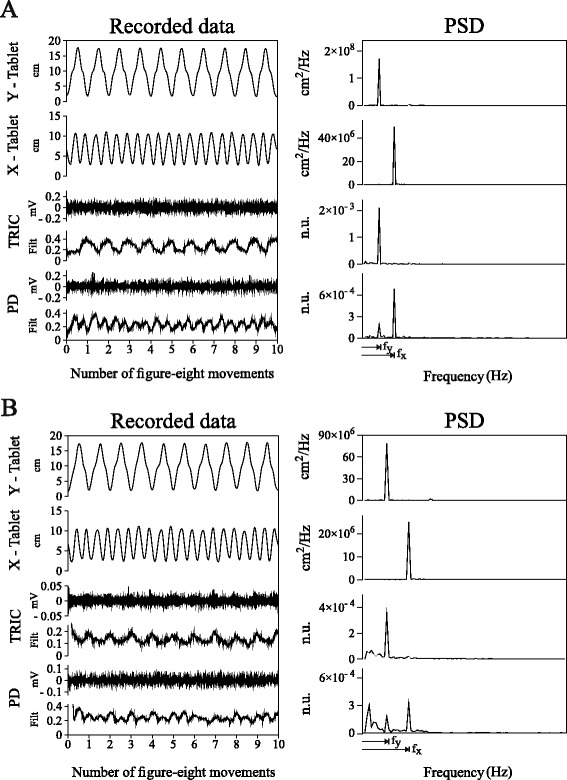
Fig. 2.

EMG-Kinematics Spectral Analysis. Panel a: Control subject (c2); Panel b: Subject with dystonia (d2). For each panel, from top to bottom: Tablet y-trajectory, Tablet x-trajectory, Triceps Brachii (TRIC) and Posterior Deltoid (PD) EMGs and non-linear envelopes (Filt) in a sequence of ten figure-eight movements represented in time (left column) and frequency (right column) domains. Note that Filt signals are normalized and dimensionless both in time and frequency domains (n.u.). fy and fx represent the subject-specific frequencies related to the vertical and horizontal components of the figure-eight
