
Fig. 4.
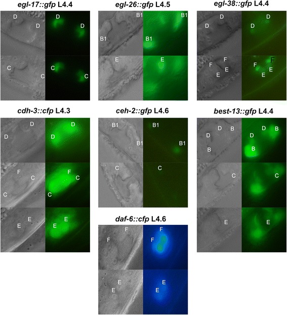
Expression of reporters in sub-stages. Each set of images shows the Nomarski image to the left and the corresponding fluorescence image to the right. The location and identity of vulval cells are indicated by "A" through "F"
Official websites use .gov
A
.gov website belongs to an official
government organization in the United States.
Secure .gov websites use HTTPS
A lock (
) or https:// means you've safely
connected to the .gov website. Share sensitive
information only on official, secure websites.
Expression of reporters in sub-stages. Each set of images shows the Nomarski image to the left and the corresponding fluorescence image to the right. The location and identity of vulval cells are indicated by "A" through "F"