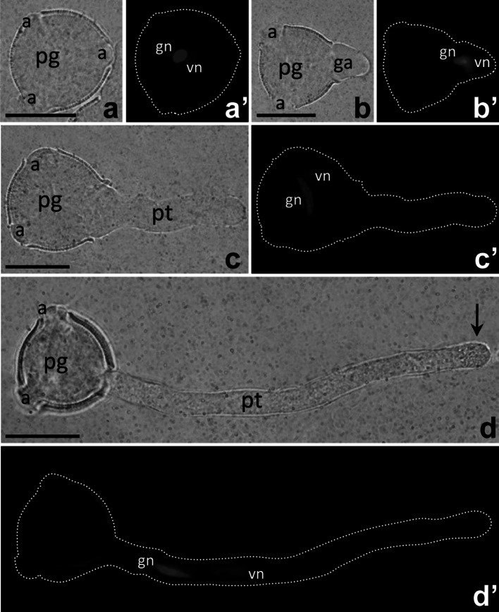
Fig. 1.
Morphology of Petunia germinating pollen (a, b) and growing pollen tubes (c, d); positions of vegetative and generative nuclei (vn and gn, respectively) in cultivated cells are visualized by DAPI staining (a′, b′, c′, d′). Arrow in d shows the pollen tube clear zone. pg pollen grain, pt pollen tube. Bars 50 μm

