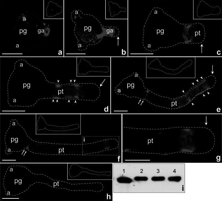
Fig. 3.
Localization of CRT protein in Petunia germinating pollen (a–c) and growing pollen tubes (d–f); g is a bigger magnification of marked region (i) in f showing the protein accumulation in the subapical zone of elongated pollen tube. Arrows in b–e, g show lack of fluorescence within the clear zone of growing pollen tubes (pt), double arrows in e, f show specific localization of CRT at the base of elongated tubes, and arrow heads in d–e show the signals localized on the peripheral cytoplasm of the growing tubes. h Negative control of immunolocalization. i Immunoblotting of crude protein extracts from Petunia anthers (2), dry pollen (3), pollen hydrated in culture medium (4), and maize anthers (1). a aperture, ga germinal aperture, pg pollen grain. Bars 50 μm

