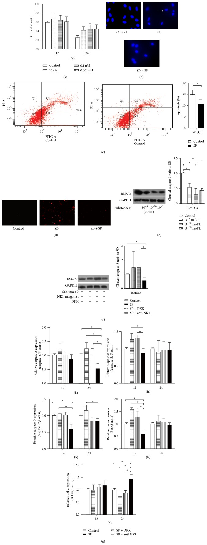
Figure 2.
Effect of SP on apoptotic changes in BMSCs. MTT assays and DAPI staining were used to quantify cell viability and to detect nuclear changes. A trend of SP reduction of SD-induced apoptotic cell death was observed from the MTT assays, although SP did not exhibit a comprehensively significant prevention in BMSCs (a). Although the concentrations ranging from 10−8 to 10−12 mol/L all exhibited a protective trend, only 10−10 mol/L SP exhibited a significant difference in comparison to the control group in BMSCs after 24 h of SD. Images of DAPI staining were taken 24 h after treatment and indicate that the nuclear condensation and integrity changes of BMSCs were reduced by SP (b). Arrows mark shrunken nuclei that indicate apoptotic cells. The flow cytometric analysis of Annexin V-FITC showed the protective trend of 10−10 mol/L SP function at 24 h SD treatment (c). Multiple images illustrate the presence of cleaved caspase-3 in BMSCs control cells and in cells subjected to 24 h SD or SD plus SP treatment. The red immunostaining of cleaved caspase-3 suggested a decrease in caspase-3-positive cells under SP treatment (d). Caspase-3 activation was quantified using Western blot analysis in the concentration manner (e) or combined with the treatment of DDK or NK-1 antagonist (f); a significant reduction in caspase-3 mRNA is indicated by qPCR (g) after 24 h of SD in BMSCs. Also, the mRNA levels of the apoptotic molecules Bcl-2, Bax, caspase-8, and caspase-9 were analyzed using qPCR in BMSCs (g). Although a clear decrease in Bax expression was not observed, the ratio of Bcl-2 to Bax was increased due to SP promotion of Bcl-2 expression. Caspase-8 expression was essentially identical between the SP group and the control group, and caspase-9 expression was reduced by SP treatment.

