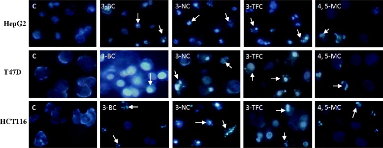
Fig. 2.

Fluorescence microscopy of the HepG2, T47D and HCT116 cells treated with the 3-BC, 3-NC, 3-TFC and 4, 5-MC (at IC50 values). Fluorescence images of the cells stained with Hoechst 33258 after 72 h. All of the four investigated chromenes induced condensation and fragmentation of the nuclei (arrows). Magnification, 200 ×
