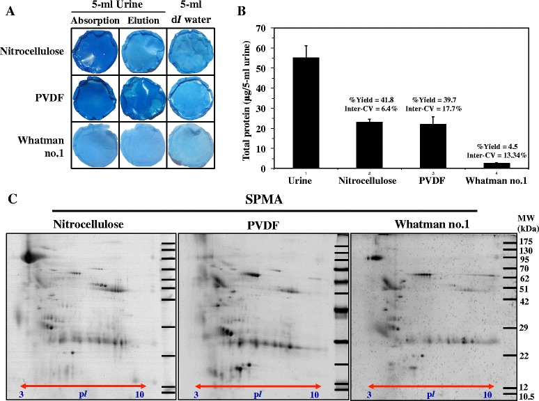
Fig. 1.

Silver Blue G-250 staining [12] showed deep blue staining of absorbed proteins on nitrocellulose and PVDF, but not Whatman no.1 membranes (a). Absorbed proteins were eluted by 2-D lysis buffer and measured by the Bradford protein assay for recovery yield and inter-CV (b). Eluted proteins were subsequently analyzed by 2-DE (50 μg/gel) with silver blue G-250 staining (c). All experiments were performed in triplicate
