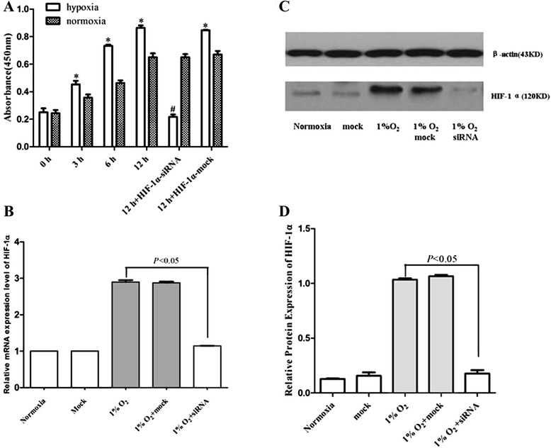
Fig. 1.

Effects of hypoxia on proliferation of AtT-20 cells. (a) AtT-20 cells were incubated under hypoxic (1% O2) and normoxia conditions for the indicated times (0, 3, 6, and 12 h). Brdu assay showed hypoxia incubation stimulated the proliferation of AtT-20 cells in a time-dependent manner compared with normoxia group (*P < 0.05 vs. normoxia groups). Cells transfected with HIF-1α-siRNA then cultured in hypoxic incubator for 12 h diminished the BrdU incorporation (#P < 0.05 vs. mock). Knock-down of AtT-20 cells with HIF-1α siRNA (10 μM) was evaluated by real-time PCR (b) and western blot (c) (P < 0.05 vs. mock). The hypoxic and normoxia incubation time is 12 h. (d) Quantification of (c). Independent experiments were repeated at least three times and similar results were obtained
